Advanced Testing
Eye exams have changed a lot over the years. Here at Dr. Lee Optometry, we pride ourselves in giving quality service with the newest advancements in eye care and imaging. Many of these tests can help diagnose and uncover many hidden eye conditions that can be difficult to catch with our basic exam.
Fundus Photography:

Fundus photography allows Dr. Lee to take a photo of your retina (the back of the eye). The retina is the surface that allows the eye to pick up light so you can see. The photo also allows the Dr. Lee to assess certain eye structures such as the macula, optic nerve and vasculature.
Being able to closely assess these structures allows for better diagnosis and early detection of certain eye diseases. Often, certain diseases such as glaucoma and age-related macular degeneration are very slow to progress and are not easily picked up by a basic exam. Fundus photography provides the ability to track changes and compare photos over time, allowing Dr. Lee to diagnose and treat the condition sooner.
Fundus photographs are especially important for people with conditions such as Diabetes, Glaucoma, Macular Degeneration, but highly recommended as a screening tool for all ages!
Optical Coherence Tomography
Optical Coherence Tomography (OCT) is a new and advanced way to view the eye. This machine allows Dr. Lee to use light rays to visualize each individual layer of the retina. Knowing exactly which layer is affected can help us better detect and diagnose ocular diseases more efficiently and provide you the best treatment possible.
Both Fundus Photography and OCT scans are typically recommended for eye exams of all ages, particularly if you have certain risk factors such as family history to certain eye diseases.

Corneal Topography

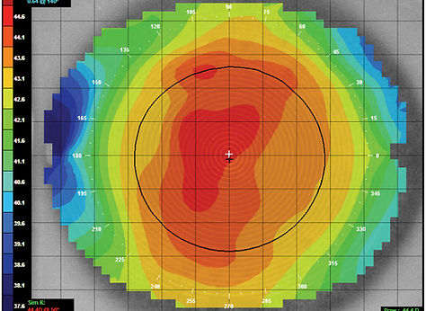
Corneal Topography is a scan of the front surface of the eye. Vision is just not about whether you are wearing glasses. A significant portion of sight can come from the shape of the front surface of the eye, the cornea.
Being able to scan the front surface of the eye allows Dr. Lee to rule out certain eye conditions that may alter it's shape. This scan is particularly important for a condition called Keratoconus, where the cornea becomes thinner causing the cornea to take on a cone-like shape instead of the normal softball shape.
This can also be used in consults for Lasik surgery or Orthokeratology.
Biometry is a relatively new scan used by optometrists. This scan sends light through the eyeball to take various measurements of the eye structures. This can be helpful in assessing cataracts for cataract surgery.
Biometry is also extremely useful in novel Myopia Management treatments. Myopia or "near-sightedness" can be due to the elongation of the eyeball. New studies and treatment methods are now available to help slow down the progression of myopia in children and Biometry is vital in assessing their effectiveness.
Biometry

Anterior OCT

Anterior OCT allows for a cross section view of the FRONT part of the eye. This is particularly important in assessing for risk of narrow angles or closed angle glaucoma.
